
Sinus Infection (Sinusitis / Rhinosinusitis)

Sinusitis is infective inflammation of the mucosal lining of one or more of the nasal sinuses. There is invariably associated changes in the lining tissue of the nose, hence the term rhinosinusitis. Swelling of the lining sinus mucosa leads to blocked drainage and ciliary malfunction which interferes with the transport of secretions. This leads to bacterial overgrowth and the production of yellow or green (purulent) mucus and an infective nasal discharge with nasal congestion. The cheek (maxillary) and front nasal bridge (anterior ethmoid) sinuses are commonly affected first, and often causes facial pressure and discomfort. An upper respiratory tract infection (URTI) and allergic rhinitis are common triggers.

Diagnostics
Nasal Rigid / Flexible Endoscopy for a detailed assessment of the inside of the nose. Skin Prick Allergy Test and RAST Allergy Test if possible rhinitis symptoms are present.
Nasal Rigid / Flexible Endoscopy
Why?
The nostrils are quite small, so even using a very bright halogen headlight and a dilator instrument (speculum) to expand the nostril, it is impossible to see any more than the front part inside the nasal cavity. The introduction of a small sterile rigid endoscope with an angled lens, or a sterile flexible endoscope whose tip can be manually rotated in various directions, allows a very detailed inspection of the whole of the nasal cavity and the site of the sinus openings. Further back the nasopharynx and Eustachian tube openings can also be examined.
How?
Cophenylcaine, a surface local anaesthetic and decongestant, is sprayed into each nostril held open by a dilator speculum. This drug is absorbed very quickly and numbs and shrinks the nasal lining. Although the introduction of an endoscope is an unusual feeling it is not unpleasant. The subsequent examination will take less than 5 minutes.
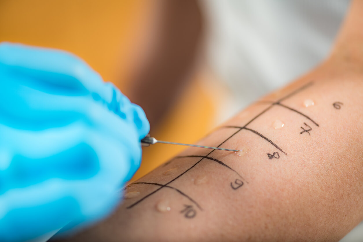
Skin Prick Allergy Test
Why?
Allergies are common and it is thought that about 1 in 3 of the population will consult a doctor at some time in their life with allergic symptoms. Many cases of rhinitis (tissue inflammation that causes persistent nasal congestion, nose bleeds, nasal itching and sneezing) can be caused by allergies.
How?
This test is simple and quick, giving results within 15-20 minutes, and is carried out by the ENT practice nurse. It is important that you do not take any form of antihistamine or steroid drug for at least 48 hours before the test as these medicines may affect the results. Allergens are introduced into the skin, usually the forearm, in such tiny amounts that testing is quite safe and can be carried out on all age groups, including babies. If you have bad eczema the test can be performed on your back.
The area to be tested is coded with a marker pen for each allergen and a drop of the solution is placed by each code. A standard concentration histamine solution is also applied to serve as a control. The skin is then pricked through the drop using the tip of a single-use sterile lancet. This can feel a little uncomfortable but should not be painful. The nurse will assess the test sites for the presence and size of redness (known as erythema) and lumps (known as wheals) after 15 minutes. The responses are compared to the control histamine solution which should always cause a reaction. The degree of reaction relative to the control indicates whether a certain level of antibodies are present which may be causing your symptoms. The wheals, which feel very much like a reaction to a nettle sting, clear within an hour for most people and any irritation can helped by applying anti-inflammatory steroid cream.
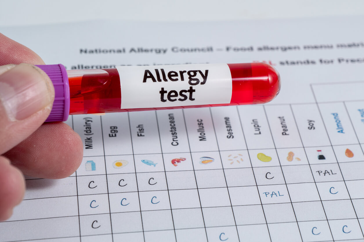
RAST Allergy Blood Test
Why?
An allergic person’s immune system produces a special type of antibody, Immunoglobulin E (IgE), to combat each specific substance to which they have an abnormal sensitivity. The RAST stands for radioallergosorbent test, which is a laboratory measure of the amount of specific IgE antibodies present in the blood.
How?
Samples of your blood are drawn from your arm at the ENT Clinic and are sent to our laboratory for analysis. The results take a few days to process but will confirm whether or not your body reacts to any substances we have tested and will also be graded to indicate the severity of the reaction. These grades range from 0-6, with 0 i
The private health insurances we work with
Harley Street ENT is recognised by a wide range of leading UK and international private health insurers, ensuring easy access to our care for patients with approved cover.


Need help and advice? Speak to an advisor
To discuss your needs in confidence or arrange a consultation, please contact our advisory team, who will be happy to assist you.